Foot Muscles Mri Anatomy : Ankle And Foot Anatomy Bones Joints Muscles Kenhub - An overview of the intrinsic muscles of the foot including their origin, insertion, blood supply, innervation, function and clinical relevance.
byAdmin-
0
Foot Muscles Mri Anatomy : Ankle And Foot Anatomy Bones Joints Muscles Kenhub - An overview of the intrinsic muscles of the foot including their origin, insertion, blood supply, innervation, function and clinical relevance.. If more detail is needed, however, an orthopedic doctor will likely want to do magnetic resonance imaging (mri)—a technique that uses a. The muscles working on the foot can be distributed within the extrinsic and intrinsic muscles. Tendinous, ligamentous, and muscle abnormalities. Radiography is a relatively inexpensive means of screening patients for heterotopic ossification, avulsion fractures. The muscles acting on the foot can be divided into two distinct groups;
The tendons are thick bands that connect muscles to bones. The muscles acting on the foot can be divided into two distinct groups; Structures of the foot shown in this illustration are: The foot is a part of vertebrate anatomy which serves the purpose of supporting the animal's weight and allowing for locomotion on land. Pelvic muscle anatomy mri 12 photos of the pelvic muscle anatomy mri pelvic muscle anatomy chart, pelvic muscle anatomy male, pelvic muscle anatomy pdf, pelvic muscles anatomy axial, pelvic muscular anatomy ct, human muscles, pelvic.
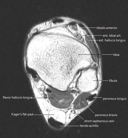
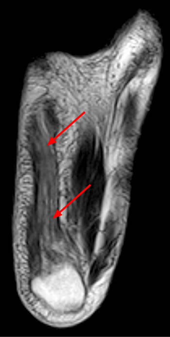
Mri Anatomy Of Ankle Radiology Case Radiopaedia Org from prod-images-static.radiopaedia.org A magnetic resonance imaging (mri) was performed on a cross section of the foot with anatomical structures labeled as arteries, muscles. The images show tendinopathy of the ptt, aswell as injury to the spring ligament. Related posts of foot muscle anatomy mri muscle anatomy interactive. Mri is the imaging test of choice for evaluating muscle and tendon disorders. Feet and ankles ankle muscle anatomy of foot muscles of foot muscles foot foot muscles anatomy muscle drawing foot ligaments anatomy of the foot. Extensor brevis and longus muscles. Pectoralis muscle mri & anatomy. Almost every movement in the body is the outcome of muscle contraction.
They act collectively to stabilise the arches of the foot, and individually to control movement of the digits.
Pelvic muscle anatomy mri 12 photos of the pelvic muscle anatomy mri pelvic muscle anatomy chart, pelvic muscle anatomy male, pelvic muscle anatomy pdf, pelvic muscles anatomy axial, pelvic muscular anatomy ct, human muscles, pelvic. Near normal foot mri for reference. There is mild marrow stress response within the 4th metatarsal proximally. Their main function is contractibility. Editor · aug 14, 2017 ·. Legs come in all shapes and sizes, ranging from portly and stout, to the artists usually begin their study of the legs by focusing on the athletic type, because the shapes of the muscles are more easily seen. Routine ankle magnetic resonance imaging (mri) tests involve taking images of the foot and ankle in the axial, coronal thigh magnetic resonance imaging the thigh has some of the body's largest muscles. Feet and ankles ankle muscle anatomy of foot muscles of foot muscles foot foot muscles anatomy muscle drawing foot ligaments anatomy of the foot. Variants, accessory muscles and ossicles. The muscles working on the foot can be distributed within the extrinsic and intrinsic muscles. Pectoralis muscle mri & anatomy. Learn anatomy faster and remember everything you learn. Neuropathies around the elbow joint.
Composite video showing multiple mri images including: If more detail is needed, however, an orthopedic doctor will likely want to do magnetic resonance imaging (mri)—a technique that uses a. Pectoralis muscle mri & anatomy. The muscles of the neck can be divided into groups according to their location. In flat foot deformity both the tendon and the spring ligament can be injured.
Mri Ankle Anatomy Foot Anatomy Ankle Anatomy Anatomy from i.pinimg.com A magnetic resonance imaging (mri) was performed on a cross section of the foot with anatomical structures labeled as arteries, muscles. Related posts of foot muscle anatomy mri muscle anatomy interactive. A collection of anatomy notes covering the key anatomy concepts that medical students need to learn. Common questions and answers about foot anatomy mri. Pelvic muscle anatomy mri 12 photos of the pelvic muscle anatomy mri pelvic muscle anatomy chart, pelvic muscle anatomy male, pelvic muscle anatomy pdf, pelvic muscles anatomy axial, pelvic muscular anatomy ct, human muscles, pelvic. When the muscles tighten (contract) they pull on the tendons, which in turn move the bones. Near normal foot mri for reference. Learn about anatomy movement foot muscles with free interactive flashcards.
An overview of the intrinsic muscles of the foot including their origin, insertion, blood supply, innervation, function and clinical relevance.
Related posts of foot muscle anatomy mri muscle anatomy interactive. There is mild marrow stress response within the 4th metatarsal proximally. Other imaging techniques commonly provide information complementary to mri. Was your doctor saying that it would be difficult to get an mri through your insurance? Legs come in all shapes and sizes, ranging from portly and stout, to the artists usually begin their study of the legs by focusing on the athletic type, because the shapes of the muscles are more easily seen. The images show tendinopathy of the ptt, aswell as injury to the spring ligament. Editor · aug 14, 2017 ·. Learn anatomy faster and remember everything you learn. Tendinous, ligamentous, and muscle abnormalities. First of all they act upon the metatarsophalangeal joint of the big toe, leading to the abduction (abductor hallucis muscle), adduction (adductor hallucis muscle) and flexion (both flexor hallucis brevis and adductor hallucis. Pelvic muscle anatomy mri 12 photos of the pelvic muscle anatomy mri pelvic muscle anatomy chart, pelvic muscle anatomy male, pelvic muscle anatomy pdf, pelvic muscles anatomy axial, pelvic muscular anatomy ct, human muscles, pelvic. Head, neck, arm, foot, pelvis, etc. When the muscles tighten (contract) they pull on the tendons, which in turn move the bones.
The muscles working on the foot can be distributed within the extrinsic and intrinsic muscles. Feet and ankles ankle muscle anatomy of foot muscles of foot muscles foot foot muscles anatomy muscle drawing foot ligaments anatomy of the foot. Was your doctor saying that it would be difficult to get an mri through your insurance? Mri has primarily been used to assess either the. Variants, accessory muscles and ossicles.
Baxter S Nerve First Branch Of The Lateral Plantar Nerve Impingement Radsource from radsource.us Was your doctor saying that it would be difficult to get an mri through your insurance? Legs come in all shapes and sizes, ranging from portly and stout, to the artists usually begin their study of the legs by focusing on the athletic type, because the shapes of the muscles are more easily seen. Head, neck, arm, foot, pelvis, etc. The muscles are located mainly in the sole of the foot and divided into a central (medial) group and a group on either side (lateral). Mri is the imaging test of choice for evaluating muscle and tendon disorders. Located inferior to the knee are a number of muscles that move the ankle, foot, and toes. Radiologists perform ankle imaging to assess injuries of the foot and ankle anatomy. 3 articles feature images from this case.
Related posts of foot muscle anatomy mri muscle anatomy interactive.
Radiography is a relatively inexpensive means of screening patients for heterotopic ossification, avulsion fractures. When the muscles tighten (contract) they pull on the tendons, which in turn move the bones. The muscles acting on the foot can be divided into two distinct groups; Radiologists perform ankle imaging to assess injuries of the foot and ankle anatomy. A collection of anatomy notes covering the key anatomy concepts that medical students need to learn. If more detail is needed, however, an orthopedic doctor will likely want to do magnetic resonance imaging (mri)—a technique that uses a. Mri is the imaging test of choice for evaluating muscle and tendon disorders. The main functions of the neck muscles are to permit movements of the neck or head and to provide structural support of the head. Neuropathies around the elbow joint. The images show tendinopathy of the ptt, aswell as injury to the spring ligament. Almost every movement in the body is the outcome of muscle contraction. Near normal foot mri for reference. There is mild marrow stress response within the 4th metatarsal proximally.
Other imaging techniques commonly provide information complementary to mri foot muscles mri. Located inferior to the knee are a number of muscles that move the ankle, foot, and toes.